CORRESPONDING ANATOMICAL TERMS
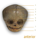
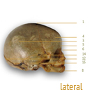
1 anterior fontanelle2 frontal bone
3 parietal eminence
4 frontal eminence
5 orbital ridge
6 glabella
7 maxilla
8 mentum
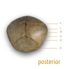
9 sagittal suture
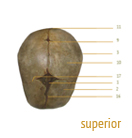
10 parietal bone
11 posterior fontanelle
12 lambdoidal suture
13 occipital bone
14 temporal suture
15 temporal bone
16 frontal suture
17 coronal suture
NOTES | 1.JONES, 2012
2. CARTA, (n.d)
2. Photos by Stef Lenk
Skull and pelvis specimens courtesy of the Scheuer collection, CAHID, University of Dundee.
THE anatomy of the human neonatal skull (click above images for full size)
Fontanelles are membranous areas that have not yet ossified in the developing cranial vault of the neonate. They allow for increased intracranial pressure during delivery and for the diameter of the skull to decrease slightly on its journey through the birth canal.1 ➡
Fontanelles are membranous areas that have not yet ossified in the developing cranial vault of the neonate. They allow for increased intracranial pressure during delivery and for the diameter of the skull to decrease slightly on its journey through the birth canal.1 ➡
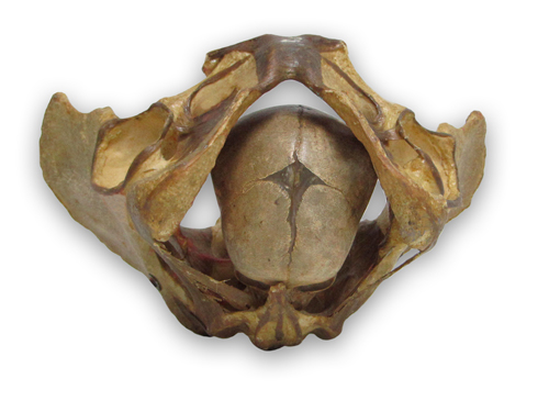
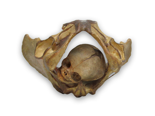
Above: the size of the neonatal skull relative to that of the female pelvis (using real specimens). Both halves of the pelvis are joined anteriorly at the pubic symphisis, a fibrocartilaginous (a mixture of fibrous tissue and cartilaginous tissues) joint that keeps the pelvis steady. During pregnancy, the symphysis pubis widens an average of 2-3 mm from the usual 4-5mm gap. The average gap is about 7.7mm. This widening of the pelvic ring helps facilitate the delivery of baby.2